Sept 2015
The paper proposes the use of short barcodes embedded in medical images to improve the speed and accuracy of image search. By generating and incorporating barcodes into DICOM files, clinicians can retrieve similar medical images and make more precise diagnoses by comparing them to retrieved cases. Additionally, image retrieval using barcodes can benefit research tasks in biomedical imaging. However, the proposal does not aim to replace feature-based classification methods commonly used in content-based image retrieval (CBIR), such as “bag of words” and “bag of features” with SVM and KNN classifiers. Barcodes are intended to supplement these existing methods and provide additional information. Barcodes can enhance the results of “bag-of-features” approaches, which typically focus on capturing the overall appearance of a scene rather than specific details of objects within the scene, such as the shape of a tumor in an MR scan. Local and region-of-interest (ROI) based barcodes are especially useful for capturing spatial information. Whether 1D or 2D barcodes are used, they are expected to yield similar results, with 1D barcodes being shorter and faster in execution.
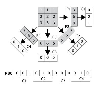
All projections (P1,P2,P3,P4) are thresholded
to generate code fragments C1,C2,C3,C4. The concatenation
of all code fragments delivers the barcode RBC.
The paper briefly reviews relevant literature on Radon transform and Local Binary Patterns (LBPs) in the context of content-based image retrieval (CBIR). The Radon transform is primarily used for representing three-dimensional objects, and various applications of the transform have been reported in the literature. These include feature detection in SAR images, extraction of invariant features for shape retrieval, and face recognition. The Trace transform, a generalization of Radon transform, has been used for detecting changes in shapes with complex textures. However, no attempt has been made to binarize Radon projections for direct use in CBIR tasks, as proposed in this paper. LBPs, introduced by Ojala et al., have been widely used in face recognition and have also found applications in CBIR. The literature on CBIR and medical CBIR is extensive, with reviews available on online systems for content-based medical image retrieval. Multiple surveys have been conducted to review recent literature in this field. Due to space limitations, the paper acknowledges that it cannot provide an exhaustive review of the vast literature on CBIR but highlights relevant works related to Radon transform and LBPs.
The Radon transform involves projecting a function f(x, y) along various angles to create a new image, R(theta, s). The projection is obtained by summing/integrating the values of f(x, y) along lines defined by each angle. The resulting image R(theta, s) is derived from the equation: s = xcos(theta) + ysin(theta). By using the Dirac delta function, the Radon transform can be expressed. To generate Radon barcodes (RBC), each projection for individual angles can be thresholded using a local threshold specific to that angle. This means applying a threshold to the projections and assembling a barcode that represents the thresholded projections. One approach for thresholding is to calculate a typical value, such as the median, based on all non-zero values in each projection. Algorithm 1 provides a description of how Radon barcodes are generated. To ensure that the barcodes have the same length, the images are normalized by resizing them.
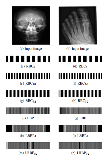
Local Binary Patterns (LBPs) have been widely used in image classification, typically by calculating their histograms as features. However, in this study, LBPs are employed differently for the purpose of comparison. They are extracted and represented as barcodes by concatenating binary vectors around each pixel. Additionally, the Galoogahi and Sim approach, known as local Radon binary patterns (LRBPs), which applies LBP on Radon transformed images, is also implemented for comparison with the proposed Radon barcodes (RBC). Figure 2 displays barcode annotations for two medical images from the IRMA dataset, showcasing different np values as indicated in lines 4 and 10 of Algorithm 1.
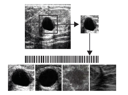
more effective intra-class retrieval. Bottom: Sample benign
(left) and malignant (right) ROIs.
In this section, the Image Retrieval in Medical Applications (IRMA) database is introduced, consisting of over 14,000 x-ray images classified into 193 categories. The images are annotated with the IRMA code, which includes four hierarchical axes representing imaging modality, body orientation, body region, and biological system examined. The IRMA dataset is divided into training and testing sets, with 12,677 images for training and 1,733 images for testing. Accuracy measurements are performed using equations to calculate the total error and the number of wrong digits in the retrieved IRMA codes. A suitability measure, denoted as _, is used to evaluate the performance based on code length, error, and mismatch. Results are presented in Table 1, indicating that the proposed Radon barcode (RBC) with 4 projections achieves the highest suitability due to its short code length. Comparisons are made with other barcode approaches, including local binary patterns (LBP) and local Radon binary patterns (LRBP). LBP shows the lowest error and wrong digits but ranks lower due to its longer code length. LRBP exhibits the highest error and wrong digits and is ranked the lowest according to the suitability measure. An additional experiment is conducted to test the implementation of ROI-based barcodes using breast ultrasound images. The success rates for correctly retrieving benign vs. malignant images using ROI-based barcodes were 7/19 for LRBP, 10/19 for LBP, and 15/19 for RBC. It should be noted that direct comparisons with previous studies using the IRMA database are not feasible due to differences in handling “don’t know” values and the absence of a classifier in this barcode-based approach.
This paper introduces the concept of barcode annotations as additional information for medical image retrieval. Radon projections are utilized and thresholded to generate barcodes. The performance of barcode-based image retrieval is evaluated using the IRMA x-ray dataset, which consists of 14,410 images for training and testing. The results suggest that Radon barcodes offer short yet informative codes that are beneficial for medical image retrieval. Future research should explore the use of barcode annotations for region-of-interest (ROI) retrieval within the same class, while inter-class retrieval can be handled by other methods such as bag-of-feature classification approaches.
Additional details: Barcode annotations for medical image retrieval: A preliminary investigation